biofilm formation protocol
Crystal Violet 1 CV1. US5928889A - Protocol for simulated natural biofilm formation - Google Patents This invention provides a methodology for controlled biofilm formation in accordance with a.
Ijms Free Full Text Expression Of The Biofilm Associated Genes In Methicillin Resistant Staphylococcus Aureus In Biofilm And Planktonic Conditions Html
Aggregation continues with the maturation of biofilm.

. Start your biofilm protocol at least 1 month before starting on antimicrobials antifungals or antibiotics. Ensure that the inlet well have about 1 mL of fresh media. This will solubilize the CV.
The 500-ml portion of autoclaved water in the stoppered flask. The development of a biofilm includes attachment of cells to a surface multiplication maturation and production of a polymeric matrix and finally microbial detachment and colonization of new surfaces. Incubate for 1015 min.
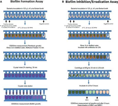
Lastly stay on a. Microtiter dish biofilm formation assay This protocol was designed to investigate biofilm formation and development by P. MeSH terms Bacteria cytology.
Biofilm Protocol Basics Conclusion. 33 CV-Stained Biofilm Quantitation 1. Aeruginosa or mutant strain overnight in a rich medium such as LB.
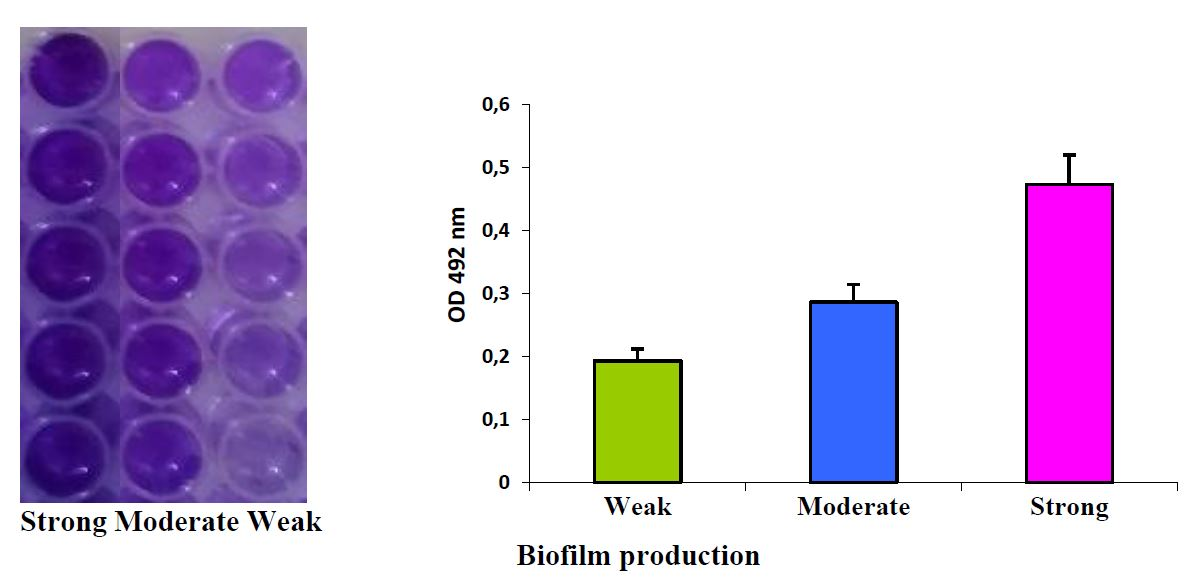
Biofilm Protocol By Kurt N. In this assay the extent of biofilm formation is measured using the dye crystal violet CV. I bf ab cw where bf is the biofilm formation ab is the od 540nm of stained attached bacteria and cw is the od 540nm of stained control wells containing bacteria-free medium only unspecific or abiotic factors kadurugamuwa et al.
Films were fixed by incubating the plates at 60C for 1 hour. The basic idea here is to recolonize your gut with a large amount of specific probiotics and nutrients. Biofilm formation is a strategy by which microorganisms survive and adapt to the involving environment particularly adverse conditions.
The main reason to avoid supplemental minerals is that most people are using Interfase Plus which is a mineral chelator. Ii bf abcw. Key points for biofilm cultivation are.
Taking minerals will just dilute the effectiveness of the product. Biofilm formation begins with attachment of bacteria to biotic surface such as host cell or abiotic surface such as prosthetic devices. Cover the tube ends with foil.
In addition these static biofilm systems allow analysis of biofilm formation with a variety of readouts including microscopy of live cells macroscopic visualization of stained bacteria and viability counts. Two methods were used to measure biofilm formation. Find a coach who is proficient in Muscle Response Testing and get your biofilm protocol basics nutritional supplements tested.
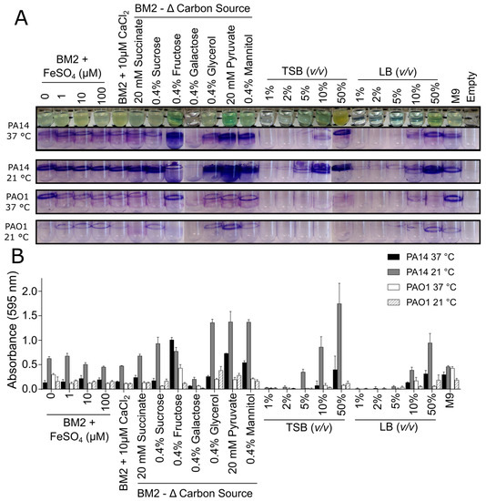
In the protocol described here we will focus on the use of this assay to study biofilm formation by the model organism Pseudomonas aeruginosa. Biofilm innoculum is usually an exhausted stationary phase culture which has been shaken vigourously overnight. Acquiring time lapse data of biofilm formation.
Rings of crystal violet around a well are not indicative of biofilm formation and should be rinsed again as excess stain will skew the results of the assay. Dispersion is started by certain conditions such as phenol-soluble modulins PSMs. This is diluted 150 or 1100 in half.
The extent of biofilm formation was determined by applying three different formulas. Step 3 is the last and my favorite step. Do not take supplemental minerals or HCl while on your biofilm protocol.
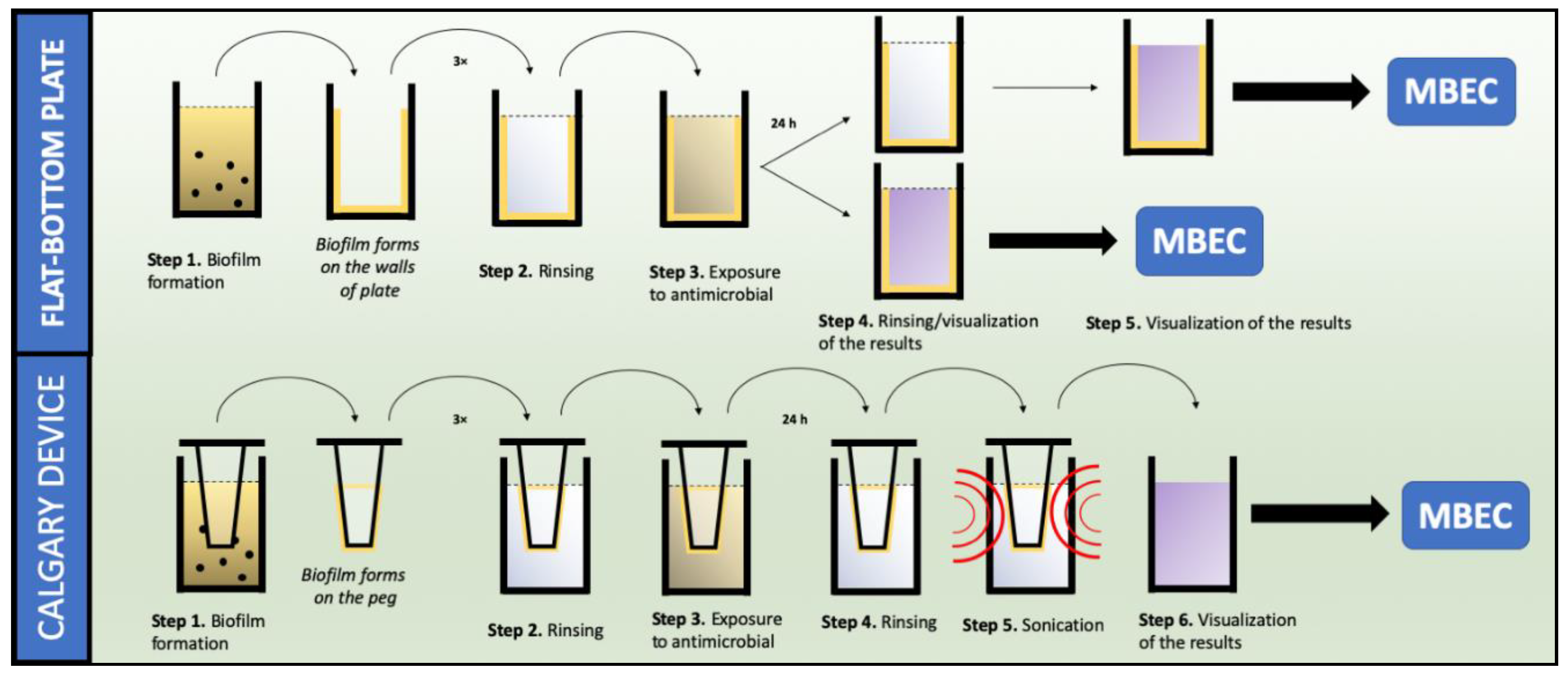
Rinse 3 times with 250 uL of sterile DI water pipetting water out 12. Biofilm development Cultivate a wild-type P. Bacterial Biofilm Protocol.
Magnesium deficiency actually contributes to bacterial virulence and biofilm formation. Used individually or in combination these assays provide useful means for the study of biofilms. Biofilm formation was evaluated by adding 200 µL of 30 acetic acid to each well after staining with 50 L of a 01 wv crystal violet solution and then measuring the OD 600 of the eluate.
Use bc 0-20 dynes plate If running experiment at 37C warm the plate and media to 37C prior to starting the experiment. Pipette up and down to assure that the stained biofilm is well solubilized and then transfer 100 μL of each sample to a new 96-well optically clear flat-bottom plate. Jain N Kohli R Cook E.
Let the plates dry completely 13. Bake microplate reader for 30minutes at 80C to adhere cells from biofilm to surface 10. The opportunistic pathogens secrete biofilm as a defense mechanism to prevent.
Leave the plate face up on the bench top overnight to dry. However the stability of biofilm does not only depend on these steps but also on the ability to inhibit other bacteria from colonizing the surface. Pipette 200 μL of 30 acetic acid solution into each well.
25th May 2020 Abhishek Dharm Singh GeneOmbio Technologies Private Limited Ammar Abou Kandil U can follow the biofilm protocol from the following article. Magnesium does not directly contribute to biofilm formation. After attachment aggregation of bacteria is started by cell-cell adhesion.
Aeruginosa and in this investigation biofilm was formed on microtiter plates 137138. Violet remaining is bound to a biofilm at the bottom of a well. As mentioned before the basic steps involved in biofilm formation are attachment of bacteria EPS secretion microcolonies formation and maturation.
Biofilm is a polysaccharide-type matrix think of a large gelatinous glob secreted not only by pathogenic and opportunistic bacteria and yeast but the normal bacteria flora in our digestive system as well. Fill separate 2-liter Erlenmeyer flasks with 15 liters water 500 ml water and 15 liters of medium appropriate for growing the biofilm of interest. Add 100uL of fresh media to the outlet well.
Your goal for this step is to Restore the gut microbiome using friendly bacteria and the building blocks your body requires in order to stabilize towards long-term health. Perfuse at 015-02 dyne for 16 to 24. Stain cells with 220uL of 01 crystal violet stain for 1 minute 11.
The remaining stain was removed by washing extensively with DI water a.

Biofilm Eradication Testing For Antimicrobial Efficacy
Biomolecules Free Full Text Critical Assessment Of Methods To Quantify Biofilm Growth And Evaluate Antibiofilm Activity Of Host Defence Peptides Html

Polystyrene Microtiter Plate Biofilm Assay Of A Pleuropneumoniae Download Scientific Diagram
Biofilm Formation Assay Kit Testpiece Dojindo Eu

Optimized Protocol For H Volcanii Immersed Liquid Biofilm Formation Download Scientific Diagram

A Cartoon Of The Methodology Presented In This Protocol Biofi Lm Download Scientific Diagram

Schematic Crystal Violet Assay On Biofilms In A Microtiter Plate Download Scientific Diagram

Biofilm Formation Assay In Pseudomonas Syringae Bio Protocol
Antibiotics Free Full Text Methods Used For The Eradication Of Staphylococcal Biofilms Html

Biofilm Formation Assay Kit Dojindo

Microtiter Dish Biofilm Formation Assay Protocol

Schematic Representation Of The Steps Involved In The Protocol For Download Scientific Diagram
Frontiers Biofilm And Planktonic Antibiotic Resistance In Patients With Acute Exacerbation Of Chronic Rhinosinusitis Cellular And Infection Microbiology

Biofilm Formation Assay Kit Dojindo

Biofilm Formation Assay Kit Testpiece Dojindo Eu
2

Biofilm Formation Assay Kit Dojindo

Microtiter Dish Biofilm Formation Assay Protocol

Crystal Violet Assay To Assess The Antibiofilm Activity Of Samples Download Scientific Diagram
0 Response to "biofilm formation protocol"
Post a Comment